+91-44-42271500
vrf@snmail.org

+91-44-42271500
vrf@snmail.org


The Radheshyam Kanoi Stem Cell Laboratory is dedicated to advancing regenerative medicine through cutting-edge research in disease modeling, tissue engineering, and cellular therapy. Our work focuses on understanding the molecular mechanisms of human diseases and developing innovative therapeutic strategies to address unmet medical needs. By leveraging advanced stem cell technologies, we aim to bridge the gap between laboratory discoveries and clinical applications. Our mission is to contribute meaningful advancements to science and improve the understanding and treatment of complex diseases.
| Name & E-Mail | Designation |
|---|---|
|
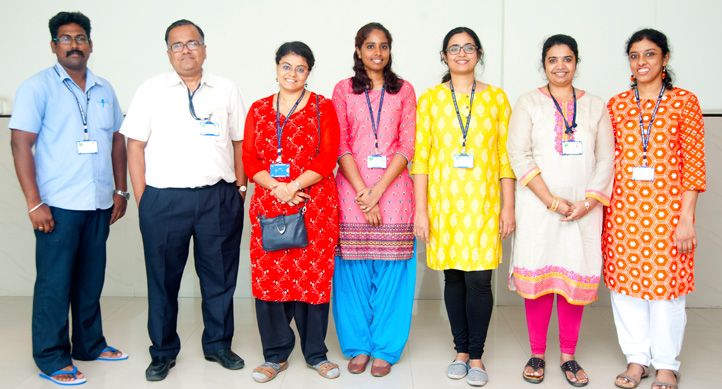
Dr Krishnakumar S
drkk@snmail.org, drkrishnakumar_2000@yahoo.com |
Professor and In charge Radheshyam Kanoi Stem Cell Laboratory |
|
Dr Nivedita Chatterjee
Email: drnc@snmail.org |
Associate Professor, Senior Principal Scientist |
|
Dr Sowmya Parameswaram
drpsowmya@snmail.org |
Associate Professor, Senior Principal Scientist |
| Ms Preethi D | Project Technical Support III |
| Mr Vinoth | Lab Attender |
Radheshyam Kanoi stem cell laboratory is dedicated to advancing research in regenerative medicine, with a strong focus on disease modeling, tissue engineering, and cellular therapy. Our studies aim to uncover the molecular mechanisms behind several human diseases and explore innovative therapeutic avenues. Below is an overview of some key research areas we are currently investigating:
Retinoblastoma, a pediatric retinal cancer, is primarily caused by germline mutations in the RB1 gene, which typically requires a second-hit mutation for tumor formation. This second-hit event, crucial for tumorigenesis, occurs in the retinain vivo but is rare in in vitro models. Most other research groups model this event by knocking out both alleles of the RB1 gene in iPSCs or ESCs, but this does not accurately reflect the biological reality. In utero, embryos with bi-allelic RB1 mutations typically do not survive, as they result in embryonic lethality. This difference presents challenges in modeling retinoblastoma using standard in vitro techniques.
To address this gap, our research uses retinoblastoma patient-derived iPSCs, specifically with a single allele (RB1+/-) knockout, to better mirror the heterozygous state seen in patients. This strategy eliminates the need for a bi-allelic knockout of the RB1 gene, which is not representative of natural embryonic development. By using single allele knockout iPSCs, we can model the early disruptions that occur in retinoblastoma patients, where the second-hit mutation plays a crucial role in tumorigenesis but is rare in early development.
We generated retinal organoids from these RB1+/- iPSCs and employed high-throughput RNA sequencing to investigate the molecular consequences of the RB1 mutation. Our results revealed significant metabolic shifts in these organoids, notably a shift from glycolysis to oxidative phosphorylation(OXPHOS), which is consistent with metabolic reprogramming often seen in cancer. These changes were accompanied by alterations in ATP and pyruvate levels, suggesting that these metabolic disruptions may be an early event that precedes other phenotypic changes seen in retinoblastoma, such as retinal cell proliferation or tumor formation.
Our model suggests that the second-hit mutation required for full tumorigenesis in retinoblastoma is rare and may not readily occur in standard in vitro settings. This rarity may point to the fact that in vivo, certain developmental factors or environmental cues in the retinal microenvironment are essential for triggering the second-hit mutation. These factors may be missing or insufficient in in vitro models, where the complex interactions of the retina and surrounding tissues are not fully replicated. This discrepancy may explain why a bi-allelic RB1 knockout in stem cell-derived retinal organoids does not automatically result in retinoblastoma-like phenotypes, as seen in more traditional models.
In summary, our findings suggest that early metabolic reprogramming—evident in the RB1+/- retinal organoids—could serve as an early biomarker of disease progression in retinoblastoma. Moreover, these results underscore the need to consider the potential absence of critical developmental signals in in vitro models, which may limit the ability of these models to fully replicate the complex, multifactorial nature of tumor initiation. Our work opens up new avenues for early intervention strategies and better understanding of the molecular basis of retinoblastoma and other RB1-related retinal diseases.
Age-related macular degeneration (AMD) is a leading cause of blindness in the elderly, primarily due to degeneration of the retinal pigment epithelium (RPE). Current therapies only address the symptoms, not the root cause. Our research investigates the potential of nano scaffolds and hypoimmunogenic induced pluripotent stem cells derived RPE in tissue engineering to develop Bruch’s membrane-mimetic substrates for AMD. This approach aims to restore RPE function and improve outcomes for AMD patients, focusing on overcoming challenges like poor RPE engraftment and retinal tissue damage.
Demyelinating diseases, such as multiple sclerosis (MS), are characterized by the degeneration of oligodendrocytes, the cells responsible for myelinating axons in the central nervous system. This loss of oligodendrocytes leads to disrupted nerve function and progressively debilitating neurological symptoms. Traditional treatments primarily aim to alleviate symptoms but do not effectively address the root cause—oligodendrocyte loss and the failure to remyelinate damaged axons.
Our research is focusing on a novel approach involving induced pluripotent stem cells (iPSCs) derived from patients to generate oligodendrocyte progenitor cells (OPCs), which are critical for the remyelination process. By reprogramming patient-specific cells, we create a source of autologous stem cells, ensuring that the therapy is ethically sound and minimizes the risk of immune rejection. These iPSCs are differentiated through optimized protocols into mature oligodendrocytes capable of myelinating axons. This approach offers a potential autologous and personalized treatmentfor demyelinating diseases by promoting remyelination.
In addition to the transplantation of iPSC-derived oligodendrocytes, our work also explores the possibility of endogenous remyelination. We have identified specific differentiation cocktails that not only enhance the ex vivo differentiation of iPSCs into oligodendrocytes but may also activate endogenous repair mechanisms in the patient’s brain. By promoting the activation and maturation of local oligodendrocyte progenitor cells (OPCs), this approach could stimulate remyelination from within, reducing the need for external cell transplantation. The cocktail of factors we have identified has the potential to both accelerate differentiation and support the maturation of oligodendrocytes, making this treatment flexible—either through transplantation of stem cell-derived oligodendrocytes or by promoting local OPC activation and differentiation.
Thus, our research represents a promising avenue for the treatment of demyelinating diseases. It leverages the power of patient-specific stem cells and could lead to personalized therapies that either supplement the body’s own regenerative potential or replace lost oligodendrocytes through transplantation, offering new hope for multiple sclerosis and other related disorders.
Retinoblastoma patients with germline RB1 mutations face an increased risk of developing osteosarcoma, particularly during adolescence. This elevated risk highlights the critical role of the RB1 gene in skeletal health and osteogenesis. We are exploring how mutations in RB1 contribute to the onset of osteosarcoma by investigating the molecular disruptions in osteogenic differentiation in mesenchymal stem cells (MSCs) from RB1 mutant and wild-type patients.
Our studies have revealed that RB1 mutant mesenchymal stem cells (OAMSCs) exhibit increased mineralization—a hallmark of osteogenesis—but without a corresponding rise in osteogenic gene expression. This suggests that RB1 mutations lead to an imbalance in normal osteogenic differentiation. Using high-throughput RNA sequencing, we identified significant disruptions in pathways related to cell proliferation, DNA repair, osteoblast differentiation, and cancer-associated pathways. These disruptions may be key factors driving the increased risk of osteosarcoma in retinoblastoma patients.
Moreover, our findings indicate a reduced expression of tumor suppressor genes and altered osteogenic markers in RB1 mutant MSCs, which could serve as early indicators of osteosarcoma. The study underscores the importance of understanding the molecular dynamics of osteogenesis in the context of RB1 mutations and highlights the need for early detection strategies in retinoblastoma patients to monitor for osteosarcoma development.
In summary, our research combines the investigation of osteosarcoma risk and osteogenic differentiation, providing valuable insights into how RB1 mutations disrupt normal bone development and contribute to an elevated risk of osteosarcoma in retinoblastoma patients. These findings may pave the way for the identification of early biomarkers for osteosarcoma and inform more targeted surveillance and therapeutic strategies for these high-risk individuals.
Retinoblastoma (RB1) mutations are traditionally linked to tumorigenesis, but recent findings have expanded their role to influencing adipogenic differentiation. Our research on orbital adipose mesenchymal stem cells (OAMSCs) derived from retinoblastoma patients has uncovered that RB1 mutations not only drive tumor formation but also contribute to increased adipogenesis, particularly promoting the differentiation of brown adipocytes. This insight is crucial as it suggests that RB1 loss could play a significant role in metabolic disorders such as obesity and Type 2 Diabetes (T2DM).
One promising therapeutic approach for T2DM is targeting adipose tissue browning, the process by which white fat transforms into energy-burning brown fat, improving insulin sensitivity and glucose metabolism. Our findings suggest that RB1 mutations may be key regulators of this process, particularly in the promotion of brown adipocyte differentiation. By understanding the molecular pathways linking RB1 loss to adipocyte differentiation, we can uncover new strategies to manage insulin resistance and treat T2DM more effectively.
In essence, our research highlights how RB1 mutations in adipogenesis could reveal novel therapeutic targets for T2DM, offering a potential pathway to enhance brown adipose tissue formation and improve metabolic health. This study lays the groundwork for developing targeted treatments for insulin resistance and other metabolic conditions, marking an exciting step forward in diabetes research and therapy.
| S.No | Items | Company |
|---|---|---|
| 1 | CO2 Incubator (2) | New Brunswick |
| 2 | Laminar flow hood (2) | Thermo Electron |
| 3 | Water bath | Nuwe |
| 4 | Cooling Centrifuge | Eppendorf |
| 5 | Microfuge | Labmate Asia |
| 6 | 37 degree Incubator-shaker | Orbitek |
| 7 | Refrigerator | Samsung |
| 8 | -20 Freezer | Hitachi |
| 9 | -80 Freezer | Thermo Electron |
| 10 | Cyclomixer | Remi/Medox |
| 11 | Fluorescent microscope with phase, digital camera | Olympus/Nikon |
| 12 | pH-meter | ThermoElectron |
| 13 | Magnetic stirrer | Scigenics Biotech |
| 14 | Cell cryopreservation liquid N2 unit | ThermoElectron |
| 15 | Platform-shaker | Scigenics Biotech |
Vision Research Foundation
No. 41 (old 18), College Road,
Chennai - 600 006, Tamil Nadu , India.
Ph No: +91-44-42271500,
+91-44-2827 1616,
vrf@snmail.org